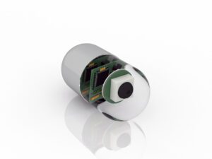
 Wireless capsule endoscopy has been very helpful in making advances in the world of endoscopy, but this work has always been limited to superficial tissue.
However, a there is a new method of pairing ultrasonography with the wireless capsule, allowing doctors to view deeper levels of tissue and therefore have a clearer view of all the tissues involved in the endoscopic procedure.
Porcine tissue has proven very valuable in researching this endoscopic procedure. The results of one such study was published in the September edition of IEEE Transactions on Medical Imaging.
In the study, tissue from the small intestine of a pig was used in vitro to determine the image resolution of the lumen wall and offers a viable resource in the fabrication of this very important device.
“Test results demonstrate that the proposed device and rotation mechanism are able to offer good image resolution (~60 μm) of the lumen wall, and they, therefore, offer a viable basis for the fabrication of a USCE device,” wrote the authors, a team of researchers from China, Scotland and the U.S.
This is yet another example of how the porcine has helped researchers make medical breakthroughs. Recently, we’ve written about their use in treating lung injuries and remedying hypertension through radiosurgical ablation of the renal nerve.
For more than 30 years, Animal Biotech has helped research teams do this kind of work by providing porcine tissues, organs, and glands. Contact us today to learn how we can assist you in your next project.


